Approaches
Magnetic Resonance Imaging and Transcranial Magnetic Stimulation (TMS) have both immense potential in the field of neuroscience:
- Structural and functional MRI allow for mapping cognitive functions to spatial and temporal features of brain activation.
- TMS holds the potential to infer on causal relationships between brain areas and cognitive functions.
- TMS and fMRI can support each other based on their respective advantages when combined.
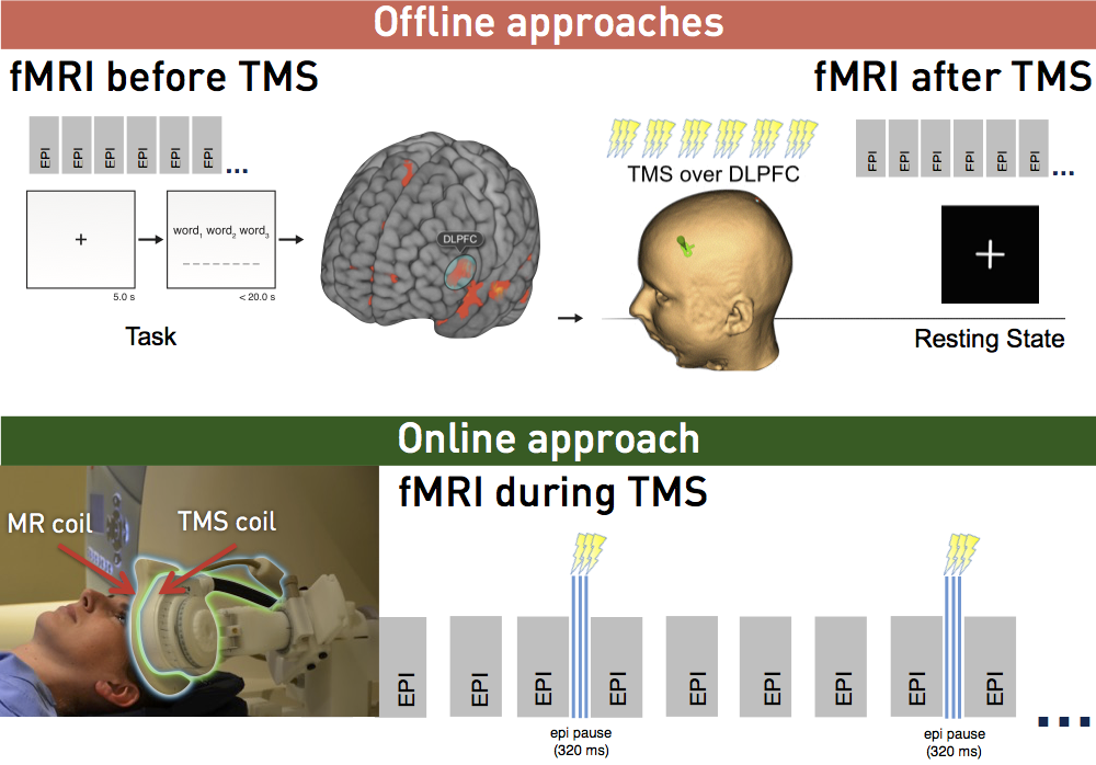
fMRI before, after and during TMS
Magnetic resonance imaging complements TMS in different ways
- Anatomical images acquired before TMS can be fed into the neuronavigation software to define the actual stimulation site.
- fMRI acquired before TMS allows for functional definition of a stimulation target.
- Offline imaging (e.g. fMRI during task or in resting-state) allows for evaluating long-term effects after TMS.
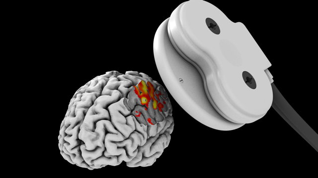
- Finally, fMRI during TMS, i.e. concurrent TMS/fMRI, allows for the direct observation of TMS-induced effects.
Concurrent TMS/fMRI
Methods for concurrent use of imaging and TMS were developed to examine TMS evoked responses in detail. This combination allows for the investigation of direct and localized neural effects as well as network effects due to TMS stimulation, yielding studies that go beyond simple observation of behavioural changes. There are, however, a number of challenges that have to be addressed for successful imaging of TMS online effects.
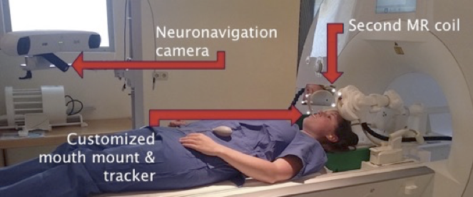
Concurrent TMS/fMRI coil array
The ideal concurrent imaging & stimulation setup has a high-density of multi-channel receive coils that are positioned directly on the subject’s scalp to maximise signal to noise ratio.
We have developed and patented a dedicated concurrent TMS/fMRI coil array.
read more