
Who we are (fMRI lab)
We are a functional neuroimaging research group at the Center for Medical Physics and Biomedical Engineering and the MR Center of Excellence of the Medical University of Vienna. Closely connected to the Vienna General Hospital, one of the largest hospitals in the world, and not too far away from the University of Vienna, the MR Center is an ideal place for our fMRI group for interdisciplinary clinical and basic biomedical research, as well as collaborations in biological psychology and cognitive science projects. Furthermore, the improvement and development of novel acquisition, data processing and analysis methods are central topics of our group.
Combination and comparison or pRF stimulation patterns
In population receptive field (pRF) mapping, we are investigating connections between the retina and the visual cortex using functional MRI. One important detail of all retinotopy experiments is the stimulation pattern presented to the subject to map the visual field. In the first place, one would suggest, that no matter in which shape the visual field is mapped has only little influence on the pRF results as long as every position is covered uniformly. However, we recently published a high-impact paper, where we show that the two most commonly used visual stimuli in pRF mapping yield systematically different results. Further, we suggest a novel combination method for the post-measurement combination of different stimuli results.
David Linhardt, Maximilian Pawloff, Allan Hummer, Michael Woletz, Martin Tik, Markus Ritter, Ursula Schmidt-Erfurth, Christian Windischberger, Combining stimulus types for improved coverage in population receptive field mapping, NeuroImage, 2021, https://doi.org/10.1016/j.neuroimage.2021.118240.
Reproducibility of Amygdala fMRI
Motivated by the recent skepticism towards the validity of fMRI tasks, particularly targeting amygdala activation, we repeated runs of an emotional paradigm and varied task instructions and technical factors. In this recent NeuroImage publication, we provide important test metrics including reliability and systematic replicability and conclude that high-resolution fMRI at 7T allows for robust mapping of amygdala activation.
Geissberger N & Tik M, Sladky R, Woletz M, Schuler AL, Willinger D, Windischberger C. Reproducibility of amygdala activation in facial emotion processing at 7T. Neuroimage. 2020 Jan 26:116585. 2020.
Woletz M, Hoffmann A, Tik M, Sladky R, Lanzenberger R, Robinson S, Windischberger C. Beware detrending: Optimal preprocessing pipeline for low-frequency fluctuation analysis. Hum Brain Mapp. 2019 Apr 1;40(5):1571-1582. 2019.
Neural correlates of the Aha-moment
We recently published a paper explaining the neural correlates and the role of dopamine circuitry when solving problems with an Aha! or Eureka-moment
(Tik M, Sladky R, Luft C, Willinger D, Hoffmann A, Banissy M, Bhattacharya J, Windischberger C. Ultrahigh-field fMRI insights on insight: Neural correlates of the Aha!-moment. Hum Brain Mapp. 2018)
International media coverage (e.g. Kurier, derStandard, Bild der Wissenschaft, Daily Mail, The Sun) has placed this paper in the Top10 most mentioned publications in Human Brain Mapping.
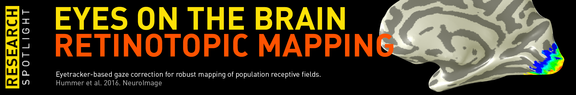
Eyes on the Brain
A new paper on artificial scotoma estimation using population Receptive Field (pRF) mapping has been recently published in NeuroImage by our group. We were able to correctly estimate small central scotoma (2.35° visual angle) on a single subject basis based on fMRI data. Investigating effects on the retinotopic map caused by artificial scotoma will help to interpret retinotopic changes observed in patient populations suffering from macular degeneration and macular holes.
(Hummer A, Ritter M, Woletz M, Ledolter A, Tik M, Dumoulin S, Windischberger C. Artificial scotoma estimation based on population receptive field mapping. NeuroImage. 2018)
Brain Connectivity in Health and Disease
Christian Windischberger was invited by the National Institute of Mental Health (NIMH) to hold a talk on Transcranial Magnetic Stimulation in Health and Disease and the related hardware developments. Our concurrent TMS/fMRI set-up allows the combination of precisely targeted neurostimulation (TMS) with high-sensitivity neuroimaging (fMRI). This promises a tool-driven revolution of TMS, allowing practitioners to individualize treatment for each patient’s need.
Watch the ▷ presentation here
Transcranial Magnetic Stimulation & Depression
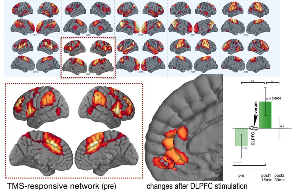
Identified network of remission (l.) and TMS influence (r.)
Martin Tik has been selected to receive a Merit Abstract Award for the 2017 OHBM Annual Meeting in Vancouver (Canada) and gave a talk on Depression and TMS as a treatment option. Depression is one of the most common psychiatric diseases and although the prognosis with gold standard treatment options is good, some patients are facing lifelong restrictions.
We were able to extract one specific fMRI resting state network with the power to discriminate full remission from early response to pharmacotherapy.
This network was found to be sensitive to repetitive Transcranial Magnetic Stimulation (rTMS) in an independent sample of healthy subjects pointing towards a distinctive mechanism of depression recovery unravelled by rTMS. We argue that rTMS over left DLPFC triggers neural processes that are comparable to those responsible for maintaining a stable healthy mental state after full remission.
TMS mechanism of action
We have published a paper on the influence of repetitive transcranial magnetic stimulation (rTMS) on resting-state connectivity. Research in this area explains the mechanism behind the beneficial effects of brain stimulation in depression (see above) and other psychiatric disorders.
(Tik M, Hoffmann A, Sladky R, Tomova L, Hummer A, Navarro de Lara L, Bukowski H, Pripfl J, Biswal B, Lamm C, Windischberger C. Towards understanding rTMS mechanism of action: Stimulation of the DLPFC Causes Network-specific Increase in Functional Connectivity. NeuroImage. 2017.